Home

Austrália múdry veľkosť ivsd echo plus veta stevard

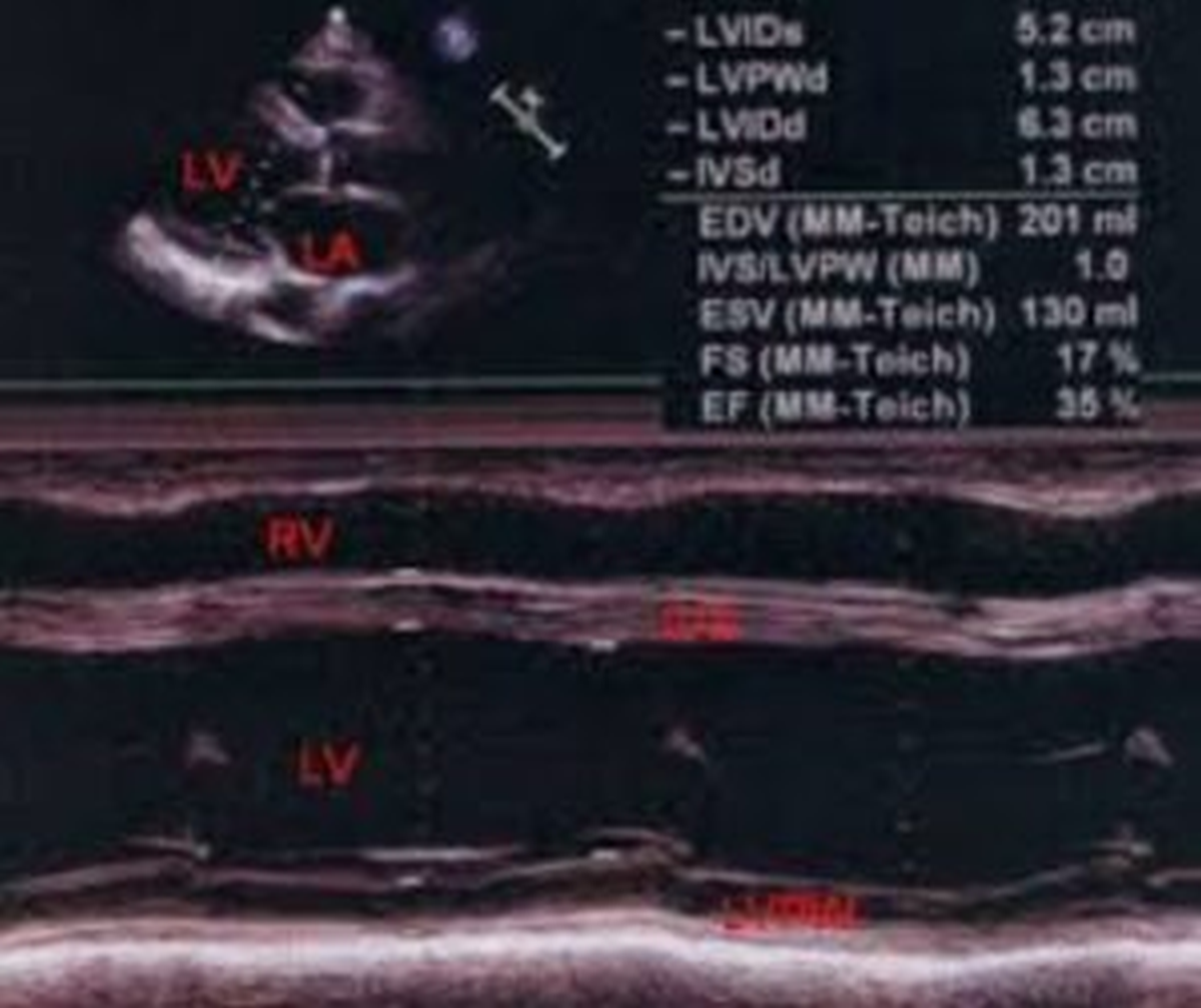
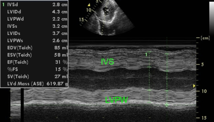
Left ventricular hypertrophy with dysfunction – M-Mode echocardiogram – All  About Cardiovascular System and Disorders
Left ventricular hypertrophy with dysfunction – M-Mode echocardiogram – All About Cardiovascular System and Disorders

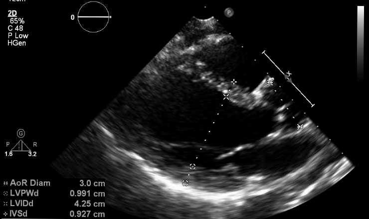
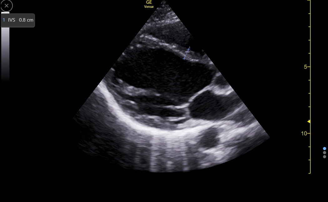
a) Example measurements of the interventricular septum thickness (IVS... |  Download Scientific Diagram
a) Example measurements of the interventricular septum thickness (IVS... | Download Scientific Diagram

Introduction to Echocardiography, Parts 1-2 | IAME

Quantitative Methods in Echocardiography—Basic Techniques | Thoracic Key

Left Ventricular Mass Assessment by 1- and 2-Dimensional Echocardiographic  Methods in Hemodialysis Patients: Changes in Left Ventricular Volume Using  Echocardiography Before and After a Hemodialysis Session - ScienceDirect
Left Ventricular Mass Assessment by 1- and 2-Dimensional Echocardiographic Methods in Hemodialysis Patients: Changes in Left Ventricular Volume Using Echocardiography Before and After a Hemodialysis Session - ScienceDirect

Measurement of left ventricular ejection fraction in parasternal long... |  Download Scientific Diagram
Measurement of left ventricular ejection fraction in parasternal long... | Download Scientific Diagram

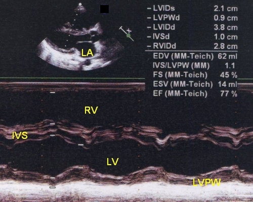
M-Mode Echocardiographic Measurements – All About Cardiovascular System and  Disorders
M-Mode Echocardiographic Measurements – All About Cardiovascular System and Disorders

What Does An Echocardiogram Show? • MyHeart

Routine Echocardiogram Protocol With Standard 2D Echo Images and Color  Doppler - YouTube
Routine Echocardiogram Protocol With Standard 2D Echo Images and Color Doppler - YouTube

Case 43: Significant Spontaneous Echo Contrast In Left Ventricle (  DCM/Severe LV Dysfunction / LVEF : 15% in M- Mode / LVEF : 10% In Simpson's  Method.... | By Interesting cases in Echocardiography | Facebook
Case 43: Significant Spontaneous Echo Contrast In Left Ventricle ( DCM/Severe LV Dysfunction / LVEF : 15% in M- Mode / LVEF : 10% In Simpson's Method.... | By Interesting cases in Echocardiography | Facebook

Echocardiography in General Practice: Cardiac Abnormalities | Clinician's  Brief
Echocardiography in General Practice: Cardiac Abnormalities | Clinician's Brief

Echocardiogram in severe pulmonary hypertension – All About Cardiovascular  System and Disorders
Echocardiogram in severe pulmonary hypertension – All About Cardiovascular System and Disorders

Quantitative Methods in Echocardiography—Basic Techniques - Echocardiography  in Pediatric and Adult Congenital Heart Disease, 2nd Ed.
Quantitative Methods in Echocardiography—Basic Techniques - Echocardiography in Pediatric and Adult Congenital Heart Disease, 2nd Ed.

Clinical and Experimental Pediatrics

M-mode echocardiogram in left ventricular dysfunction – All About  Cardiovascular System and Disorders
M-mode echocardiogram in left ventricular dysfunction – All About Cardiovascular System and Disorders

Echocardiography - Wikipedia

Small animal cardiac ultrasound measurements | IMV imaging

Long axis view of echocardiography demonstrates IVS hypertrophy and... |  Download Scientific Diagram
Long axis view of echocardiography demonstrates IVS hypertrophy and... | Download Scientific Diagram

LV Size and Function

M-Mode echocardiogram in LV dysfunction – All About Cardiovascular System  and Disorders
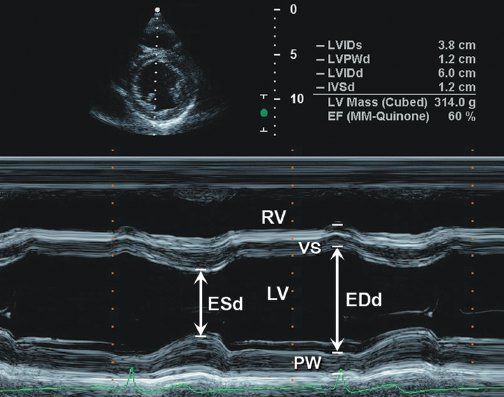
M-Mode echocardiogram in LV dysfunction – All About Cardiovascular System and Disorders
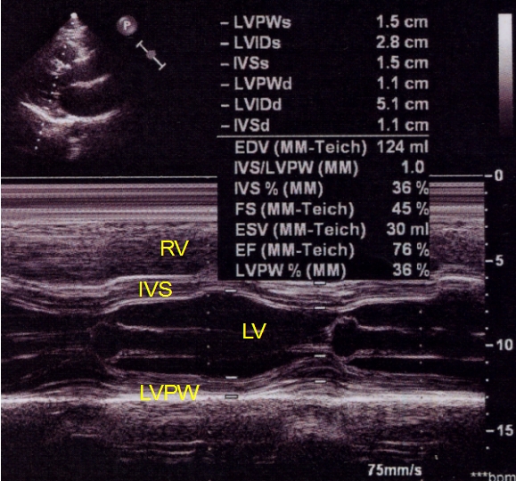
Concentric left ventricular hypertrophy by echocardiography. IVSd,... |  Download Scientific Diagram
Concentric left ventricular hypertrophy by echocardiography. IVSd,... | Download Scientific Diagram